
C. T. Li1, M. Wick1, and N.G.
Marriott2
1The Ohio State University Department of
Animal Sciences
2Virginia Polytechnic Institute and State
University
Lipid oxidation is a major contributor to flavor deterioration in meat products. A modified peroxide value (mPV) method was compared to the 2-thiobarbaturic acid (TBA) test as methods of analyzing the lipid oxidation state in subcutaneous lamb fat obtained from lambs fed either 15 IU or 300 IU supplemental a-tocopherol (vitamin E). The mPV and TBA analyses both demonstrated no significant difference (P > 0.05) in the lipid-oxidation state of the fat from four different loin sections (n = 20). Both mPV and TBA methods demonstrated virtually identical differences (P < 0.05) in the lipid-oxidation state of the fat derived from lambs fed two different levels of vitamin E. These results demonstrate the efficacy of employing mPV to monitor the lipid-oxidation in animal fat. The low temperatures inherent to the mPV method vs. the TBA method, along with mild extraction methods and speed of obtaining results, reduces the potential of causing spurious autoxidation or generating substances that are capable of interfering with the assay. Both mPV and TBA methods indicate that the level of dietary intake of vitamin E significantly affects the lipid oxidation-state of subcutaneous fat antemortem as well as seven days postmortem and, thus, the shelf life of lamb fat.
Flavor is the trait responsible for consumer preferences for meat and meat products. Water-soluble compounds in the lean portion of muscle impart meat taste while the lipids contribute the flavors characteristic of lamb species (Horstein and Crowe, 1963). Lipid oxidation during prolonged storage or short-term exposure to high temperatures is often associated with "off flavors," "warmed over flavor," "rancid," and "stale" characteristics in mutton which result in product degradation and reduced case-life of an otherwise nutritious protein source.
One of the most important causes of meat food flavor deterioration is lipid oxidation, which affects fatty acids in general and polyunsaturated fatty acids in particular (Gray, 1978; Allen and Allen, 1981; and Fennema, 1993). Postmortem factors can influence lipid oxidation and decrease the shelf life of meat products due to the initiation of peroxidation (Vercellotti et al., 1992). Oxidation of fatty acids in animal tissue starts to occur almost instantly after slaughter (Gray and Pearson, 1994).
Autoxidation of lipids is carried out by a free- radical chain reaction (Gray, 1978; Allen and Hamilton, 1983; Rojarho and Sofos, 1993). The initial step in this reaction is the generation of transitory hydroperoxides, which degrade into malonaldehyde (MA) and several other reactive compounds. Due to their unstable state, peroxides start their decomposition and form a series of secondary products, such as aldehydes, acids, and ketones, that produce undesirable rancid flavors (Shahidi, 1994).
The most common chemical measurement of lipid oxidation in muscle foods is the thiobarbaturic acid (TBA) assay, based on the reaction of MA and TBA generating a TBA-MA complex with an absorbance maximum at 530 nm (Patton and Kurtz, 1951). Although widely employed, this method has limitations; the most significant being that both MA and TBA react with other substances present in meat and the extracts of meat. These substances produce aldehydic lipid oxidation products that can react with TBA. In addition, the nonspecificity of the assay is due to interfering compounds that react with TBA such as sugars, ascorbic acid, and nonenzymatic browning products (Decker et al., 1998). Another potential drawback to the TBA method is that MA is often bound to proteins and the conditions for the optimal release of MA is often hard to determine. These forms vary from one sample to the next and require different hydrolytic conditions to release the MA. Also, it is difficult to release all of the MA from meat protein without employing strong acids and heat that adversely affect stability of the TBA-MA complex (Draper et al., 1986).
A comparison of a modified peroxide value (mPV) and TBA (2-thiobarbituric acid) as methods for analyzing the lipid oxidation state of the lipids in the subcutaneous fat layer obtained from lambs fed two different levels of the lipid soluble antioxidant a-tocopherol (vitamin E) is reported here. These results demonstrate that mPV is a rapid, low-cost, simple, and reproducible method to monitor the lipid oxidation state in animal fat.
Comparison of TBA and mPV
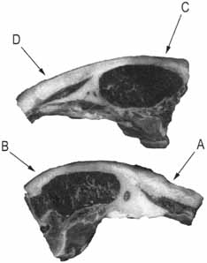
For comparative statistical analysis of mPV and TBA methods, two samples of subcutaneous fat were removed from the right side of the loin and two samples from the left side of the loin. The lamb was obtained from The Ohio State University Meat Lab, Columbus, Ohio (Figure 1). The lamb had been fed an all-concentrate diet, offered ad libitum, formulated to provide 16% crude protein and 15 IU (NRC; Control) of supplemental vitamin E per kg of diet dry matter. It should be noted that lambs are normally fed 15 IU supplemental vitamin E per kg of diet dry matter and as such are considered control lambs. Carcass fabrication was conducted one-day post-slaughter.
Subcutaneous fat was removed, wrapped in a meat tray with polyvinyl-chloride oxygen-permeable film, and stored at 39.2 ± 3.6°F (4 ± 2°C) prior to analysis by both TBA and mPV.
|
| Figure 1. Sampling locations of subcutaneous fat from lamb loins. Right loin: A, B; Left loin: C, D. |
Analysis of Lipid Oxidation State in Lambs Fed Two Vitamin E Supplements
A 2 (levels of vitamin E) x 4 (days of storage) factorial comparison of the lipid oxidation state in the subcutaneous loin fat in lambs fed two different levels of supplemental vitamin E was performed by employing both TBA and mPV. Twelve lambs were obtained from The Ohio State University’s Ohio Agricultural Research and Development Center (OARDC) campus at Wooster, Ohio, where they had been fed an all-concentrate diet, offered ad libitum, formulated to provide 16% crude protein and either the control 15 IU supplemental vitamin E, recommend by National Research Council, or 300 IU supplemental vitamin E per kg of diet dry matter, seven days before slaughter. Carcass fabrication was conducted seven days post-slaughter. Subcutaneous fat from the loin area was removed, wrapped in a Styrofoam meat tray with polyvinyl-chloride oxygen-permeable film, and stored at 39.2 ± 3.6°F (4 ± 2°C) from 1 to 11 days post-slaughter.
2-Thiobarbituric Acid (TBA) Assay
The degree of lipid oxidation in this experiment was determined by the TBA method described by Pensel (1990). Briefly, 5.0-grams of unrendered lamb fat were placed into a coded polyethylene stomacher bag. An additional empty stomacher bag was prepared as a blank. Then 50-mL of 39.2 ± 3.6°F (4 ± 2°C) 20% trichloroacetic acid (Fisher Scientific, Fair Lawn, N.J.) in 1.6% of m-phosphoric acid (Fisher Scientific, Fair Lawn, N.J.) solution was immediately added to each stomacher bag. Samples were blended in a Seward Laboratory blender (Tekmar Co., Cincinnati, Ohio) for 2 min. Then 50-mL of 39.2 ± 3.6°F (4 ± 2°C) distilled water were added to each bag for a second blending for 30 s. The slurry was filtered through a Whatman No. 1 filter. Five mL of freshly prepared 0.02 M 4, 6-dihydroxypyrimidine-2-thiol (Sigma Chemical Co., St. Louis, Mo.) was added to each tube and mixed for 4—5 s. Tubes were stored in the dark for 15 hours to develop the color. The color was measured by a Gilford Response UV-VIS Spectrophotometer (Ciba Corning Diagnostic Co., Oberlin, Ohio) at a wavelength of 530 nm.
A standard curve for the indirect determination of K was generated by reacting TBA with 1,1,3,3 -tetra-ethoxypropane (TEP) (Sigma Chemical Co., St. Louis, Mo.). A K value of 9.242 was obtained (n = 20, R2 = 0.996). The TBA of each sample was determined employing the following formula:
TBA mg MA/g = K x O.D530nm
Modified Peroxide Value (mPV)
The PV method of analysis was modified from the AOAC (1990) procedure. Briefly, a 50 g sample of unrendered subcutaneous loin fat each was ground in a Waring lab blender (Waring Products Division, Dynamic Cooperation of America, New Hartford, Conn.) for 20 to 30 s and extracted with 30 mL ice cold (3:2 v/v) acetic acid:chloroform. The extraction was vigorously swirled to distribute the sample and reagents. After the samples were dissolved in the acetic acid:chloroform mixture, 0.5 mL of saturated potassium iodine (KI) (83.2 g solid KI/40 mL H2O) were added and mixed vigorously. Subsequently, 30 mL deionized water were added and the solution mixed thoroughly. Color of the upper aqueous layer ranged from pale yellow to bright yellow, with the lower organic layer remaining white. The mixture was allowed to stand for 5 to 10 min. at room temperature then titrated with 0.01 M Na2S2O3 (Sigma Chemical, Fair Lawn, N.J.) gradually with vigorous shaking. During the titration, 0.5 mL of starch indicator (Starch 1% with chloroform 0.3%, Lab Chem., Inc., Pittsburgh, Pa.) was added. Color of the upper aqueous layer ranged from light purple to dark purple, and the lower organic layer remained white to gray. If the color of the lower organic layer remained yellow, the sample was vigorously swirled and allowed to stand for an additional 10 min. The end-point of titration was established when the color of the upper aqueous layer disappeared. The mPV was calculated employing the following formula:
mPV = (S)(N)(1000) W
Where: mPV = Modified Peroxide Value
(meq oxygen/kg fat)S = mL Na2S2O3 N = Normality of Na2S2O3 (0.01 N) W = g of fat
Statistical analysis was completed by statistical software SAS 7.0 for Windows® (SAS Institute, Inc., Cary, N.C.). Two-Way General Linear Models (GLM) were used to analyze the results.
Comparison of TBA and mPV Methodologies
The results reported in Table 1 indicate that both TBA and mPV methods detected no differences in the lipid oxidation state at four locations in the subcutaneous loin fat (P > 0.05, n = 20). In addition, results also demonstrate the consistency and repeatability of both TBA and mPV. In addition, these data also indicate that lipids in the subcutaneous fat surrounding the loin from lambs fabricated within 24 hours of slaughter exhibit a very low oxidation state.
Table 1. Lipid Oxidation at Different Locations of Lamb Fat by TBA and mPV Methods.1 | ||
|---|---|---|
| Locations N= 5/location |
mPV (meq/kg) |
TBA (µg/g) |
| A | 2.48a | 0.150a |
| B | 2.36a | 0.133a |
| C | 2.04a | 0.138a |
| D | 1.96a | 0.143a |
|
1Means followed by the same superscripts within columns indicate that the values are not different (P > 0.05). Right loin: A, B; Left loin: C, D. TBA = 2-thiobarbaturic acid and mPV = modified peroxide value. | ||
The comparative advantages of mPV in contrast to TBA as a method of analyzing the lipid oxidation state of lamb fat are summarized in Table 2. The ability of mPV to detect the oxidation of mono unsaturated fatty acids suggests that this method is more accurate in determining the oxidation state of all the fatty acids in the sample in contrast to the traditional TBA methodology. The potential for further autoxidation of the sample is minimized in the mPV method due to the use of non-acidic and ice-cold conditions during sample preparation and extraction. Additionally, the potential to compare the oxidation state in fat derived from different muscles is more appropriate with mPV than traditional TBA due to the fact that mPV detects only the degree of peroxide formation in the unconjugated fatty-acid fraction of the animal fat (Decker et al., 1998). Finally, final color development for mPV is almost instantaneous while color development for the traditional TBA method requires at least 15 hours. This greatly shortens the "turn around" time for analysis of the lipid oxidation state of lamb fat by mPV versus the traditional TBA method.
Table 2. Comparative Analysis Between 2-Thiobarbaturic Acid (TBA) and the Modified Peroxide Value (mPV). | ||
|---|---|---|
| mPV | TBA | |
| Temperature of sample preparation (°C) | 7 ± 2 | 25-100 |
| Sample autoxidation potential | Low | High |
| Analysis time | 2 h | 18—24 h |
| Acidic conditions | No | Yes |
| Specificity for peroxides | Yes | No |
| Detection of oxidized mono and unsaturated fatty acids | Yes | No |
|
Potential to compare oxidation between muscles with different fatty acid composition |
Appropriate | Inappropriate |
Use of TBA and mPV to Determine Lipid Oxidation State of Fat from Lambs Fed Different Vitamin E Regimens
Both mPV and TBA methods demonstrate an increase in the lipid oxidation state in loin area subcutaneous fat obtained from lambs fed on both the control and 300 IU supplemental dietary vitamin E (P < 0.05). Both methods demonstrate similar lipid oxidation states of the fat from animals fed on both regimens until eleven days post slaughter. The greatest increase in 11-day lipid oxidation was observed in the fat from lambs fed 15 IU (control animals) by both methods. There was no difference in the mPV or TBA of fat obtained from animals fed 300 IU vitamin E on days 1, 7, or 9, suggesting that high dietary vitamin E treatment retards the rate of lipid oxidation to a greater extent than control levels of dietary vitamin E treatment. In addition, lambs fed 15 IU vitamin E had higher initial mPV and TBA values than those fed 300 IU vitamin E (Table 3). Furthermore, mPVs significantly increased between day 9 and 11 (P < 0.05), indicating that increased dietary vitamin E treatment reduces the rate of lipid oxidation more than control dietary vitamin E intake, thus increasing the usable shelf life of lamb fat.
Table 3. Effects of Two Different Levels of Dietary Vitamin E on the Modified Peroxide Value (mPV) and 2-Thiobarbaturic Acid (TBA) Values of Lamb Fat During Storage.1 | ||||
|---|---|---|---|---|
| Day Post Slaughter |
mPV (meq/kg) |
TBA (µg/g) | ||
| Control | 300 IU | Control | 300 IU | |
| 1 | 1.90a | 1.03a | 0.167a | 0.084a |
| 7 | 2.08ab | 1.09a | 0.179ab | 0.090a |
| 9 | 2.33b | 1.11a | 0.193b | 0.101a |
| 11 | 3.50c | 1.94b | 0.254c | 0.125b |
| 1IU: International Unit. Total observation, N = 96. Means followed by different superscripts within columns indicate statistical differences are significantly different (P < 0.05) | ||||
Previous research indicated that supplemental dietary vitamin E reduces lipid oxidation state of rendered fat from lamb meat. The rate of lipid oxidation is controlled by various antemortem factors. Composition of the diet is one of the most effective ways of inhibiting lipid oxidation in animal fat (Wulf et al., 1995). In this study, lambs fed 300 IU of vitamin E apparently had lower mPVs than those fed 15 IU. These results suggest that higher levels of dietary vitamin E contribute to lower mPVs and less oxidation of subcutaneous lamb fat. In addition, increased levels and longer duration of dietary vitamin E contributed to a greater inhibition of lipid oxidation in the subcutaneous loin fat in lambs.
The mPV method reported here reproducibly analyzes the lipid oxidation state of fatty acids derived from animal tissue. The reaction time for the entire analysis is less than three hours. In contrast, the TBA method reported here is time consuming and incapable of detecting oxidized mono-unsaturated fatty acids.
The TBA method is primarily based on determining the concentration of a TBA-MA complex, a secondary oxidative product of fatty acid oxidation. However, MA is not present in all oxidized systems. In addition, the TBA method is not sensitive to the oxidation state of mono- or di-unsaturated fatty acid derivatives (Nawar, 1996). Gray (1978) reported no color development by TBA method for linoleate, a mono-unsaturated fatty acid, even though the peroxide value had already reached to 2,000 (Gray, 1978). Interaction of MA with available amino groups in meat components has been reported to affect the results of TBA analysis (Shahidi, 1994).
Lipid oxidation determinations are normally performed on animal fat samples more than seven days post slaughter. This is the first report of the lipid oxidation state determination on fresh (< 24 hours), unrendered, fabricated subcutaneous lamb fat. Both mPV and TBA yielded similar results, however; mPV is more rapid, simpler, more reproducible, costs less, and uses unrendered fat. These aspects of the mPV method could have a potentially great economic contribution for the meat industry.
Research supported by funds awarded to
M. Wick from OARDC #617216-A265.
Allen, C. E. and E. Allen. 1981. Some lipid characteristics and interactions in muscle foods: a review. Food Technol. 35:253.
Allen, J. C. and R. J. Hamilton. 1983. Rancidity in Foods. Applied Science Publishers, London.
AOAC. 1990. Official Methods of Analysis of the Association of Official Analytical Chemists. Ch. 4, pp. 956. Association of Official Analytical Chemists, Inc. Arlington, Va.
Decker, E. A., Chan, W. K. M., and Faustman, C. 1998. TBA as an index of oxidative rancidity in muscle foods. Proc. 51st Ann. Recip. Meat Conf. p. 66. American Meat Science Assoc., Kansas City, Mo.
Draper, H. H., McGirr, L. G., and Hadley, M. 1986. The metabolism of malonaldehyde. Lipids 21(4):305.
Fennema, O. R. 1993. Quimica de 10s alimentos. Ed. Acribia, Zaragoza, Spain.
Gray, J. I. 1978. Measurement of lipid oxidation: A review. J. Am. Oil Chem. Soc. 55:539—546.
Gray, J. I. and Pearson, A. M. 1994. Lipid-derived off-flavor in meat-formation and inhibition. In: Flavor of Meat and Meat Products. 1st Ed. pp. 117-139. Chapman & Hall, London, U.K.
Hornstein, I. and Crowe, P. F. 1963. Meat flavors: Lambs. J. Agric. Food Chem. 11:147.
Nawar, W. W. 1996. Lipids. In: Food Chemistry. 3rd Ed. pp. 226, 255—273. Marcel Dekker, Inc., New York, N.Y.
Patton, S. and Kurtz, G. W. 1951. 2-Thiobarbituric acid as a reagent for detecting milk fat oxidation. J. Dairy Sci. 34(6):699.
Pensel, N. A. 1990. Influence of experimental conditions on porcine muscle and its effect on oxidation. Masters Thesis. Department of Animal Sciences, The Ohio State University, Columbus, Ohio.
Raharjo, S. and Sofos, J. N. 1993. Methodology for measuring malonaldehyde as a product of lipid peroxidation in muscle tissues: A review. Meat Sci. 35:145.
SAS. 1998. SAS User’s Guide: Statistics and statistical analysis system, version 7. SAS Institute, Inc., Cary, N.C.
Shahidi, F. 1994. Assessment of lipid oxidation and off-flavor development in meat and meat products. In: Flavor of Meat and Meat Products. pp. 247-266. Chapman & Hall, London, U.K.
Vercellotti, J. R., St. Angelo, A. J., and Spanier, A. M. 1992. Lipid oxidation in foods: An overview. In: Lipid Oxidation in Food. pp. 1-11. American Chemical Society, Washington, D.C.
Witte, V. C., Krause, G. F. , and Bailey, M. E. 1970. A new extraction method for determining 2-Thiobarbituric acid values of pork and beef during storage. J. Food Sci. 35:582—585.
Wulf, D. M., Morgan, J. B., Sanders, S. K., Tatum, J. D., Smith, G. C., and Williams, S. 1995. Effects of dietary supplementation of vitamin E on storage and caselife properties of lamb retail cuts. J. Anim. Sci. 73:399—405.
1 For more information, contact at: The Ohio State
University 230A Plumb Hall, 2027 Coffey Road, Columbus, OH 43210, (614)
292-7516, Fax (614) 292-7116; email:wick.13@osu.edu
2 Department
of Food Science and Technology, Virginia Polytechnic Institute and State
University, Blacksburg, Virginia 24061